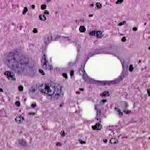
 | Scrapie: Lesions in the gray matter of the brain of a sheep with scrapie: (A) typical spongiform change in neurons; (B) spongiform change and astrocytic hypertrophy and hyperplasia. A, hematoxylin and eosin stain; B, glial fibrillar acid protein (GFAP) stain. Magnification x500. [Micrograph courtesy of Dr. R. Higgins, School of Veterinary Medicine, University of California, Davis] Download TIF for Image 1 |