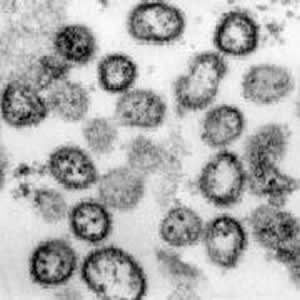
 | Lassa virus, the causative agent of Lassa fever, an important hemorrhagic disease of West Africa. Thin section of virions in a space between cells—Lassa virus buds from the surface membrane of cells where it is then free to invade other nearby cells and is free to enter the bloodstream. Magnification approximately x55,000. Micrograph from F. A. Murphy, University of Texas Medical Branch, Galveston, Texas. Download TIF |
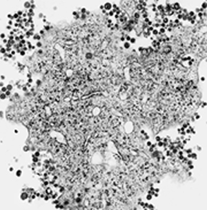
 | Lassa virus, low magnification image of an infected Vero cell in culture. Lassa virus buds from the surface membrane of cells where it is then free to invade other nearby cells and is free to enter the bloodstream. Many in vitro studies of arenaviruses employ techniques such as zonal ultracentrifugation that select for median-sized virions, say virions about 110nm in diameter, leaving the impression that the arenaviruses are quite unifrom in size. However, as shown here, they really are quite variable in size and pleomorphic in outline. Magnification approximately x15,000.
Micrograph from F. A. Murphy, University of Texas Medical Branch, Galveston, Texas. |