Thin section electron microscopy of infected cell culture and negative contrast electron microscopy of virus from cell culture. |
 | Ebola virus (image 1), diagnostic specimen from the first passage in Vero cells of a specimen from a human patient—this image is from the first isolation and visualization of Ebola virus, 1976. This image has been "borrowed" so often for various public uses that many people think that all Ebola virions look just like this—indeed, in the film Outbreak every virion seen looked just like this. In fact, Ebola virions are extremely varied in appearance -- they are flexible filaments with a consistent diameter of 80 nm (nanometers), but they vary greatly in length (although their genome length is constant) and degree of twisting. Negatively stained virions. Magnification: approximately x60,000. Micrograph from F. A. Murphy, University of Texas Medical Branch, Galveston, Texas. Download TIF |
| | |
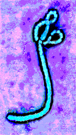 | Ebola virus (image 1 colorized 1), diagnostic specimen from the first passage in Vero cells of a specimen from a human patient—this image is from the first isolation and visualization of Ebola virus, 1976. This image has been "borrowed" so often for various public uses that many people think that all Ebola virions look just like this—indeed, in the film Outbreak every virion seen looked just like this. In fact, Ebola virions are extremely varied in appearance — they are flexible filaments with a consistent diameter of 80 nm (nanometers), but they vary greatly in length (although their genome length is constant) and degree of twisting. Negatively stained virions. Magnification: approximately x60,000.
Micrograph from F. A. Murphy, University of Texas Medical Branch, Galveston, Texas. Download JPG Only |
| | |
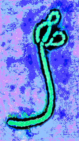 | Ebola virus (image 1 colorized 2), diagnostic specimen from the first passage in Vero cells of a specimen from a human patient—this image is from the first isolation and visualization of Ebola virus, 1976. This image has been "borrowed" so often for various public uses that many people think that all Ebola virions look just like this—indeed, in the film Outbreak every virion seen looked just like this. In fact, Ebola virions are extremely varied in appearance — they are flexible filaments with a consistent diameter of 80 nm (nanometers), but they vary greatly in length (although their genome length is constant) and degree of twisting. Negatively stained virions. Magnification: approximately x60,000. Micrograph from F. A. Murphy, University of Texas Medical Branch, Galveston, Texas. Download JPG Only |
| | |
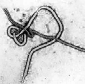 | Ebola virions (image 2), diagnostic specimen from the first passage in Vero cells of a specimen from a human patient — this image is from the first isolation and visualization of Ebola virus, 1976. In this case, some of the filamentous virions are fused together, end-to-end, giving the appearance of a "bowl of spaghetti." Negatively stained virions. Magnification: approximately x40,000. Micrograph from F. A. Murphy, University of Texas Medical Branch, Galveston, Texas. Download TIF |
| | |
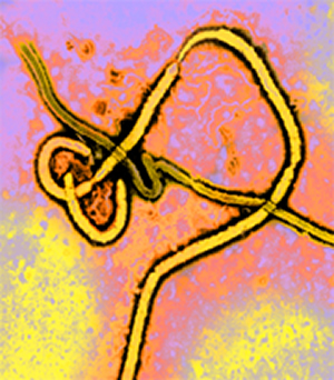 | Ebola virions (image 2 colorized 1), diagnostic specimen from the first passage in Vero cells of a specimen from a human patient — this image is from the first isolation and visualization of Ebola virus, 1976. In this case, some of the filamentous virions are fused together, end-to-end, giving the appearance of a "bowl of spaghetti." Negatively stained virions. Magnification: approximately x40,000. Micrograph from F. A. Murphy, University of Texas Medical Branch, Galveston, Texas. Download Large JPG |
| | |
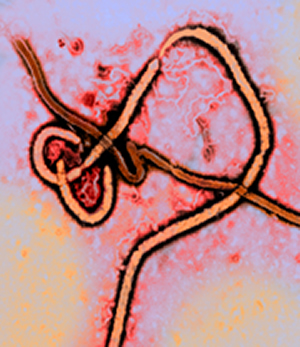 | Ebola virions (image 2 colorized 2), diagnostic specimen from the first passage in Vero cells of a specimen from a human patient — this image is from the first isolation and visualization of Ebola virus, 1976. In this case, some of the filamentous virions are fused together, end-to-end, giving the appearance of a "bowl of spaghetti." Negatively stained virions. Magnification: approximately x40,000. Micrograph from F. A. Murphy, University of Texas Medical Branch, Galveston, Texas. Download TIF |