Global web ALERT button

Eastern equine encephalitis virus

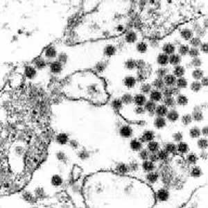
Eastern equine encephalitis virus -  Thin section electron microscopy of infected cell culture.

Eastern_equine_encephalitis_virus

Eastern equine encephalitis virus. This is an ultra-thin section of a Vero cell culture infected for 24 hours. Virions have accumulated in the space between cells as a result of budding from the surface membrane of infected cells. In this infection very large numbers of the 60 nm (nanometer) spherical virions are produced quickly and as quickly the cells are destroyed. Magnification approximately x70,000.

Micrograph from F. A. Murphy, University of Texas Medical Branch, Galveston, Texas.

Download TIF


Eastern_equine_encephalitis_virus_Aedes_salivary_gland_thumbnailEastern equine encephalitis virus, in mosquito salivary gland. This is an ultra-thin section of the salivary gland of an Aedes triseriatus (Say) mosquito, 21 days after infection, showing large numbers of virions within the lumen of an upstream divereticulum of the salivary space. In this infection large numbers of the 60 nm (nanometer) spherical virions crowd the salivary space, ready to be transmitted to the next vertebrate host when the mosquito seeks a blood meal. Magnification approximately x70,000.

Micrograph from F. A. Murphy, University of Texas Medical Branch, Galveston, Texas.

Download TIF