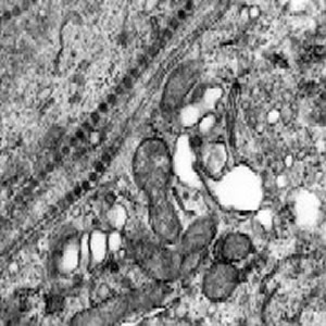
Thin section electron microscopy of infected mouse brain.

This is an ultra-thin section of the brain of a mouse infected with Bunyamwera virus (family Bunyaviridae, genus Bunyavirus) at six days post-infection when the mouse was moribund. Virions have accumulated in the normally narrow spaces between neurons as a result of budding from the intracytoplasmic (Golgi) membranes and exocytosis via membrane fusion. Some virions are still located within the lumen of Golgi vesicles. Magnification approximately x40,000.
Micrograph from F. A. Murphy, University of Texas Medical Branch, Galveston, Texas.
Download TIF